NUCLEAR MEDICINE IN NEUROIMAGING

PETCT and TRODAT Scan for Brain

Alzheimer's Disease

Dementia is a complex syndrome affecting areas of cognition, problem-solving, memory amongst many others. Often times there is a significant overlap of clinical presentation of various dementia disorders.
Functional imaging can objectively diagnose, predict and differentiate various dementia disorders at a much higher sensitivity and specificity then conventional imaging. Nuclear medicine offers PET and SPECT imaging technology for the same.
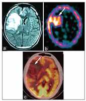
This functional imaging modality is based on the utilisation of caused by the neurons. Areas showing abnormal function show a reduction in glucose utilisation and hence can be seen as an area of hypometabolism on the brain PET imaging.
Since funcåonal changes proceed the development of structural changes, brain PET can pick abnormalities earlier then morphologic abnormalities

Why PET CT in DEMENTIA? Assessment of subjects with clinical Mild Cogni ve Impairment( MCI)-

Amongst the current available imaging modalies FDG brain PET is the most sensive technique for evaluaon of Alzheimer’s disease.

Paent’s clinical MCI progresses to develop frank Alzheimer’s disease while a small subgroup stays stable.
Subjects with clinical MCI and an abnormal abrain PET are 18 mes more likely to develop frank Alzheimer’s disease in due course of me when compared with those subjects with normal Brain PET

Abnormal brain PET in AD
Normal brain PET

PET CT in Other Demena Disorders:

  • Diffuse Lewy Body demena 
  • FrontoTemporal Demena and its subtypes
  • CorcoBasal Degeneraon

Movement Disorders: MRI Brain vs TRODAT scan

Movement disorders, typically Parkinson’s disease becomes overtly evident aer over 50% neuronal loss on MRI Scan. Tc-TRODAT scan, which images the dopaminergic system, can objecvely demonstrate dopaminergic deficit (which leads to Parkinson’s disease) much earlier during the course of disease, being the only imaging modality capable of doing so.
It can also reliably differenate drug-induced Parkinsonism versus Parkinson’s disease.

Parkinson’s disease

Normal
TRODAT scan

PET CT + TRODAT scan - Parkinson-Plus Syndrome

Combinaon of brain PET and Tc-TRODAT scan is valuable in the evaluaon of Parkinson’s plus syndromes. These scans along with clinical neurologic evaluaon provide the highest diagnosc yield in evaluaon of movement disorders with demena.

Coregistered PET MRI Brain Scan Evaluaon of brain tumors:

Tc-GHA SPECT study: Based on the principle of blood-brain barrier breakage, this study reliably differenates a post- treatment inflammaon versus a recurrence with the highest sensivity. Representave images from *literature:

Reference DOI: 10.4103/0972-3919.147525

Figure 8: A 30-year-old male with right frontal grade II astrocytoma, primarily treated with radiotherapy, presented with severe headache. Magnec resonance imaging (a) was posive for residual/recurrent tumor (arrow). 99m-techneumglucoheptonate single photon emission computed tomography (b) was also strongly posive for recurrent/residual tumor (arrow). However, on 18F-fluorodeoxyglucose positron emission tomography/computed tomography (c) the recurrent/residual lesion was hypometabolic (arrow)

Evaluaon of Refractory Seizures:

Over 1/4th of the paents with epilepsy disorder evolve into refractory epilepsy. These paents can potenally be treated successfully with neurosurgical resecon if the paent has a focal epileptogenic abnormality that is safe to remove. FDG brain PET can idenfy the seizure focus.

Specific ulity of FDG Brain PET in seizures is:

  •  Idenfy focus when MRI is normal or discordant with EEG findings
  • To guide site of invasive subdural electrode placement
  • Lateralise and idenfy epileptogenic focus in cases of bilateral abnormalies on MRI
  • Infants, where incomplete myelinaon limits evaluaon with MRI imaging
  • To exclude contralateral abnormalies in paents being planned for hemispherectomy
At Infinity Medical Centre, we have a worked towards creang a mulmodality imaging protocol that involves interictal FDG brain PET and MRI brain as one single study. This enables a perfect correlaon between structural informaon by the MRI and funconal informaon by the brain PET, enabling precise and more valuable informaon

022 6917 2400 / 022 6917 2424
80921 21212

infopet@infinitymedicalcentre.co.in

Next to T. B. Hospital Main Gate, Koch’s House, Jerbai Wadia Road, Sewri, Mumbai - 400 015.

Copyright © 2024 IMC All Right Reserved.